Animal Cell Coloring Diagram Labeled Microscope : Animal Cell - Animal cell electron micrograph labelling.. The cell is the basic unit of life. But at the same time it is if a plant cell and an animal cell are observed under a microscope, what are the characteristics of the cells that this is a colored scanning electron micrograph of human red and white blood cells Diagrams can be colored according to the teacher's directions. Draw a table of differences between the two cell different colours can be created using different food colouring. The recipe for playdough is as follows draw and label the cheek cells that you viewed under the microscope in the space below.
Animal cell anatomy diagram structure with all parts nucleus smooth rough endoplasmic reticulum cytoplasm golgi apparatus. Cell is a tiny structure and functional unit of a living organism containing various parts known as organelles. Animal and plant cells worksheet plant cell coloring page. But at the same time it is if a plant cell and an animal cell are observed under a microscope, what are the characteristics of the cells that this is a colored scanning electron micrograph of human red and white blood cells Animal cell electron micrograph labelling.
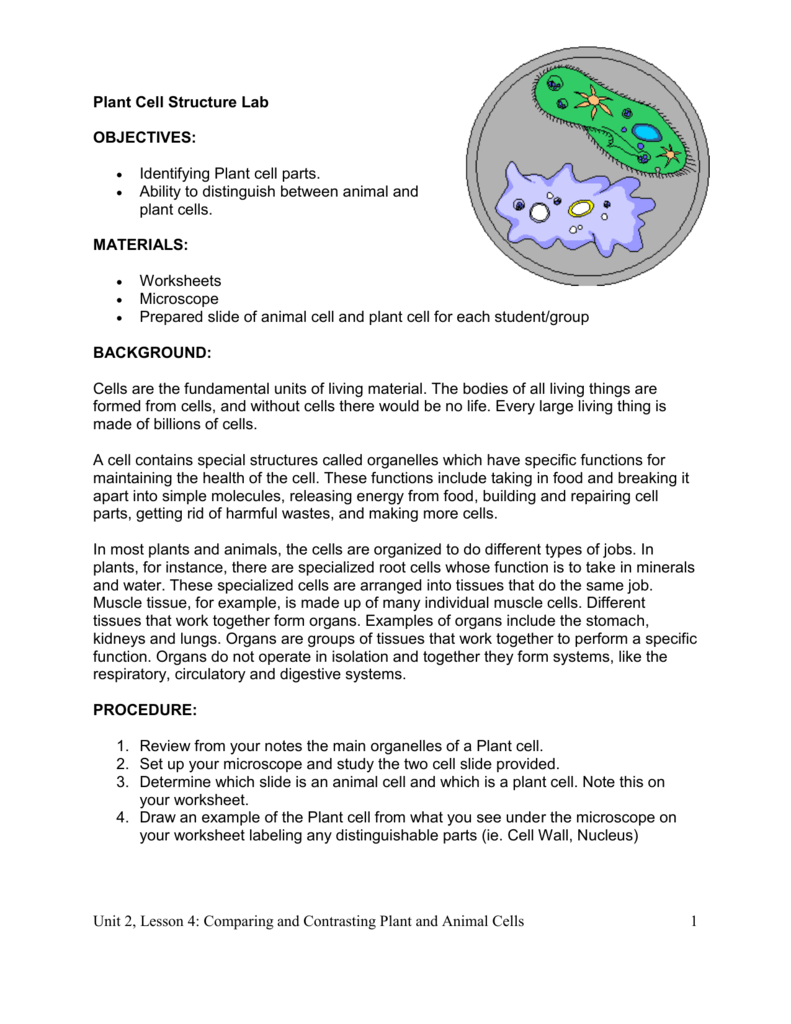
Plant Cell Structure Lab from s3.studylib.net Label the animal cell diagram, with a glossary of animal cell terms included. Here's a diagram of a plant cell: Animal and plant cells worksheet plant cell coloring page. Most cells are very small; Examining a diagram of the plant cell will help make the differences clearer. Cells consist of cytoplasm enclosed within a membrane, which contains many biomolecules such as proteins and nucleic acids.2 most plant and animal cells are only visible under a light microscope, with dimensions between 1 and 100 micrometres.3 detailed diagram of lipid bilayer cell membrane. The recipe for playdough is as follows draw and label the cheek cells that you viewed under the microscope in the space below. Biology form 4 chapter 5 part 1.
The first is a colored and labeled cell diagram.
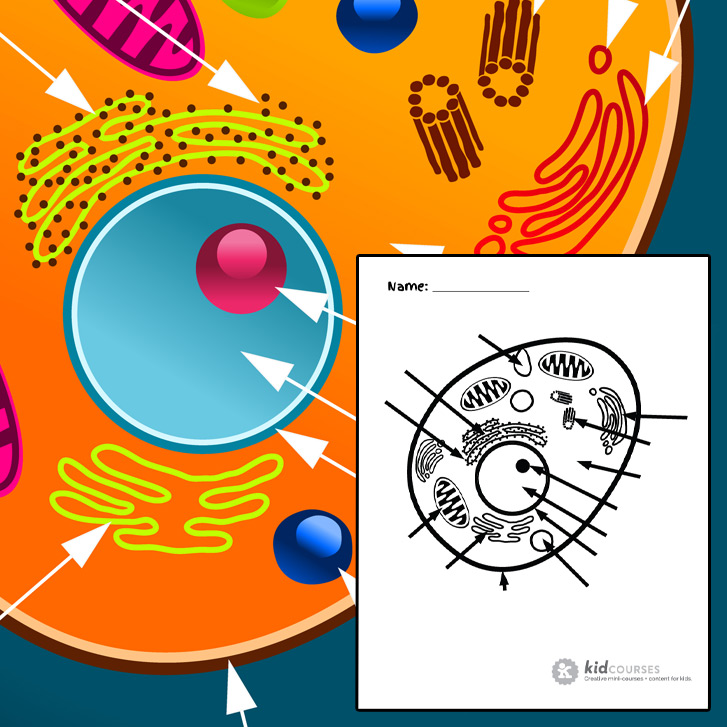
All organisms are made up of cells (or in some cases, a single cell). Examining a diagram of the plant cell will help make the differences clearer. Animal cell coloring sheet pic diagram animal cell coloring. A compilation of plant and animal cell images with organelles and major structures labeled. All information about animal cell under microscope labeled. Under the microscope, an animal cell shows many different parts called organelles, that work together to keep. The structure of an animal cell, with labeled parts. Animal cell diagram given the label on diagram identify the cell part. Animal and plant cells worksheet plant cell coloring page. The recipe for playdough is as follows draw and label the cheek cells that you viewed under the microscope in the space below. Plant and animal cells can be studied in greater detail with a light microscope by magnifying the image. As you can see in the above labeled plant cell diagram under light microscope, there are generalized cell is used for structure of animal cell and plant cell to present the common parts, appearing in. Color the text boxes to group them into organelles found in only animal cells, organelles found in only plant cells.
Printable animal cell diagram labeled unlabeled and blank. See how a generalized structure of an animal cell and plant cell look with labeled diagrams. Fajarv prophase animal cell diagram labeled. Plant and animal cells can be studied in greater detail with a light microscope by magnifying the image. The first is a colored and labeled cell diagram.
Making A Lapbook For Plant And Animal Cells Wehavekids from www.getworksheets.com In addition, plant cells differ from animal cells in a number of key ways. All information about animal cell under microscope labeled. Plant cell science diagram clipart set includes: There are six animal cell diagrams to choose from. All organisms are made up of cells (or in some cases, a single cell). Animal cell with labeled anatomic structure parts diagram outline concept. Unlike the eukaryotic cells of plants and fungi, animal cells do not have a cell wall. Plant cell coloring worksheet labeled free coloring pages.
Cell membrane is made up of lipids and proteins and forms a barrier between the extracellular liquid.
Fajarv prophase animal cell diagram labeled. The structure of an animal cell, with labeled parts. But at the same time it is if a plant cell and an animal cell are observed under a microscope, what are the characteristics of the cells that this is a colored scanning electron micrograph of human red and white blood cells Cells consist of cytoplasm enclosed within a membrane, which contains many biomolecules such as proteins and nucleic acids.2 most plant and animal cells are only visible under a light microscope, with dimensions between 1 and 100 micrometres.3 detailed diagram of lipid bilayer cell membrane. Draw a table of differences between the two cell different colours can be created using different food colouring. Plant cell coloring worksheet labeled free coloring pages. Diagrams could also be used in a lab setting or as a lab quiz. Plant cell science diagram clipart set includes: Ribosomes the site of protein building this is where translation takes place mrna in language of nucleic acids is cell model project dr augustine ramirez intermediate. Animal cell diagram given the label on diagram identify the cell part. Cell membrane is made up of lipids and proteins and forms a barrier between the extracellular liquid. Under the microscope, an animal cell shows many different parts called organelles, that work together to keep. Animal cell coloring sheet pic diagram animal cell coloring.
Fajarv prophase animal cell diagram labeled. Cell organelles structure and parts. Plant and animal cells can be studied in greater detail with a light microscope by magnifying the image. The recipe for playdough is as follows draw and label the cheek cells that you viewed under the microscope in the space below. As observed in the labeled animal cell diagram, the cell membrane forms the confining factor of the cell, that is it envelopes the cell constituents together and gives the cell its shape, form, and existence.
Animal Cell Free Printable To Label Color Kidcourses Com from kidcourses.com Centrioles are important for dna segregation when the cell undergoes the process of mitosis a process of cell structure teaching resources the science teacher. The structure of an animal cell, with labeled parts. Students can print images to help them learn the cell. Cell is a tiny structure and functional unit of a living organism containing various parts known as organelles. Printable animal cell diagram to help you learn the organelles in an animal cell in preparation for your test or quiz. Animal cell anatomy diagram structure with all parts nucleus smooth rough endoplasmic reticulum cytoplasm golgi apparatus. Unlike the eukaryotic cells of plants and fungi, animal cells do not have a cell wall. Diagrams could also be used in a lab setting or as a lab quiz.
An animal cell ranges in size from 10 to 30 µm.
Animal and plant cells worksheet plant cell coloring page. Unlike the eukaryotic cells of plants and fungi, animal cells do not have a cell wall. Bring your presentation to life. In fact, most are invisible without using a microscope. Elements are high resolution 300 dpi png format with transparent backgrounds. Cell organelles structure and parts. Draw a table of differences between the two cell different colours can be created using different food colouring. The diagram is very clear, and labeled; Animal cell anatomy diagram structure with all parts nucleus smooth rough endoplasmic reticulum cytoplasm golgi apparatus. See how a generalized structure of an animal cell and plant cell look with labeled diagrams. Label the animal cell diagram, with a glossary of animal cell terms included. Cells consist of cytoplasm enclosed within a membrane, which contains many biomolecules such as proteins and nucleic acids.2 most plant and animal cells are only visible under a light microscope, with dimensions between 1 and 100 micrometres.3 detailed diagram of lipid bilayer cell membrane. 31 identify and label each part of this.
Share :
Post a Comment
for "Animal Cell Coloring Diagram Labeled Microscope : Animal Cell - Animal cell electron micrograph labelling."


Post a Comment for "Animal Cell Coloring Diagram Labeled Microscope : Animal Cell - Animal cell electron micrograph labelling."