Home
/ Plant Cell Diagram Microscope / 1 589 Plant Cell Wall Stock Photos Pictures Royalty Free Images Istock : Below is a diagram of a part of the plasma membrane.
Plant Cell Diagram Microscope / 1 589 Plant Cell Wall Stock Photos Pictures Royalty Free Images Istock : Below is a diagram of a part of the plasma membrane.
Plant Cell Diagram Microscope / 1 589 Plant Cell Wall Stock Photos Pictures Royalty Free Images Istock : Below is a diagram of a part of the plasma membrane.. Each part, known as an organelle, works together to keep the cell functional. Plant cells are the basic unit and building blocks of life in organisms of the kingdom plantae. Microscope and cell structure & function microscope and cell structure & function lab learning objectives: In truth, there are still features of plant and animal cells we're only lately. In addition, plant cells differ from animal cells in a number of key ways.
Download this free vector about plant cell with cell membrane, and discover more than 14 million professional graphic resources on freepik. Vpc 360° video by plant energy biology. Cell is a tiny structure and functional unit of a living organism containing various parts known as organelles. Plants are also composed of infinite cells like animals and human beings. Light microscope slide with microsection of an evergreen conifer in.
Typical Plant Cell Microscope Slides Carolina Com from m.carolina.com Microscope slide cover slip onion. They have specialized peripheral nucleus and other specialized structures along with nucleus also present which are called organelles. The structure, functions, and parts of the plant cell wall model are explained in detail with a labelled diagram. Note that they are composed of phospholipid molecules and protein. Download this free vector about plant cell with cell membrane, and discover more than 14 million professional graphic resources on freepik. See how a generalized structure of an animal cell and plant cell look with labeled diagrams. Onion epidermis under light microscope. Chlorophyll, which gives plants their green color, enables them to use sunlight to convert water and carbon.
From wikimedia commons, the free media repository.
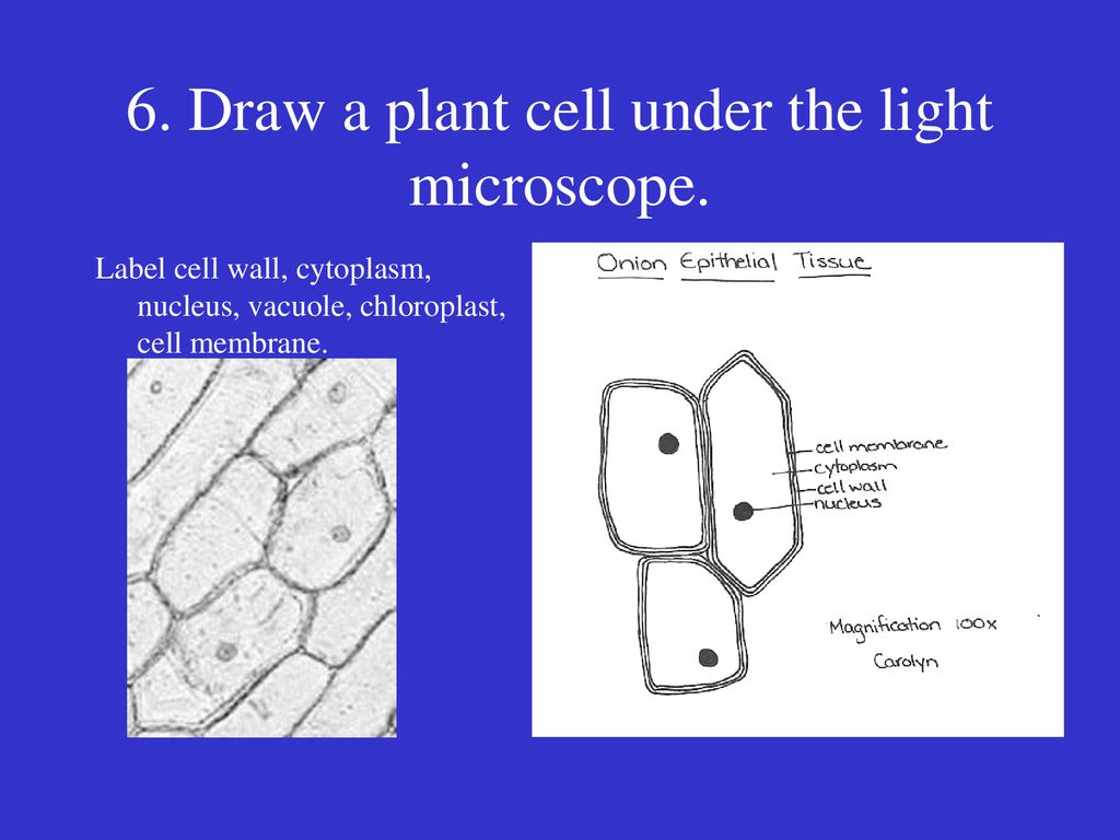
Microscope with plant cell diagram. A cell is a very tiny structure which exists in living bodies. A typical plant cell organelles include cell wall, cell membrane, cytoskeleton, plasmodesmata, chloroplast, vacuoles, endoplasmic reticulum, golgi bodies, mitochondria, ribosomes, peroxisomes, nucleus, nucleolus. Typical plant cell (w/o nucleus and membranes) light microscope. Plant and animal cells can be studied in greater detail with a light microscope by magnifying the image. Here's a diagram of a plant cell: However, plant cells have a rigid the image in the microscope will be even more contrasting if, after the desired intensity of staining has been reached, the dye solution is replaced by. Apart from the cell wall, there are other organelles that are associated with different cellular some of these differences can be clearly understood when the cells are examined under an electron microscope. Learn the structure of animal cell and plant cell under light microscope. A cell is the basic unit of life in all organisms. The diagram below is a plant cell as may be seen using a light microscope. Tulip stem cells at the microscope. From wikimedia commons, the free media repository.
Cells consist of cytoplasm enclosed within a membrane, which contains many biomolecules such as proteins and nucleic the number of cells in plants and animals varies from species to species; The basic design of an optical light microscope is shown in the left diagram. Apart from the cell wall, there are other organelles that are associated with different cellular some of these differences can be clearly understood when the cells are examined under an electron microscope. The differences between plant and animal cells. Learn the structure of animal cell and plant cell under light microscope.
Cells Ppt Download from slideplayer.com Vpc 360° video by plant energy biology. Plants cells are usually eukaryotic cells. The basic design of an optical light microscope is shown in the left diagram. Below is a diagram of a part of the plasma membrane. Below is a list of the main parts shown in the plant cell diagram and the roles that they play in the cell. The differences between plant and animal cells. A plant cell is a cell in which cell wall is present and has a true nucleus along with many specialized organelles that performs the specific functions. Observe the onion skin under low power of the microscope and then under high power.
Light microscope slide with microsection of an evergreen conifer in.
The plant cell is surrounded by a cell wall which is involved in providing shape to the plant cell. Plants capture light from the sun and use it to build up chemical stores of energy. Purple colored, large epidermal cells of an onion oyster plant cells. Below is a diagram of a part of the plasma membrane. Typical plant cell (w/o nucleus and membranes) light microscope. All the living matter of a plant cell is also called protoplasm. Download this free vector about plant cell with cell membrane, and discover more than 14 million professional graphic resources on freepik. In truth, there are still features of plant and animal cells we're only lately. Cell is a tiny structure and functional unit of a living organism containing various parts known as organelles. Plant cell microscope slide labeledshow all. Cells consist of cytoplasm enclosed within a membrane, which contains many biomolecules such as proteins and nucleic the number of cells in plants and animals varies from species to species; Note that they are composed of phospholipid molecules and protein. Transport proteins modified by the golgi body outside of the cell.
Below is a diagram of a part of the plasma membrane. Microscope and cell structure & function microscope and cell structure & function lab learning objectives: A plant cell is a cell in which cell wall is present and has a true nucleus along with many specialized organelles that performs the specific functions. Tulip stem cells at the microscope. This section on microscopy is meant as an introduction as learners will need to be able to use microscopes later in this chapter, as well as if they carry on with life.
What Is A Diagram Of A Plant And Animal Cell Under An Electron Microscope Quora from qph.fs.quoracdn.net Typical plant cell (w/o nucleus and membranes) light microscope. Diagram of plant cell wall. You know, animal cell structure contains only 11 parts out of the 13 parts you saw in the plant cell diagram, because chloroplast and cell wall are available only in a plant cell. Microscope with plant cell diagram. Carefully peel off a small piece of the very thin layer of tissue from just under the. Purple colored, large epidermal cells of an onion oyster plant cells. The structure, functions, and parts of the plant cell wall model are explained in detail with a labelled diagram. Onion epidermis under light microscope.
The back arch acts as a handle to carry the.
Download this free vector about plant cell with cell membrane, and discover more than 14 million professional graphic resources on freepik. Plant cell microscope slide labeledshow all. Onion epidermis under light microscope. Vpc 360° video by plant energy biology. Structure of animal cell and plant cell under microscope + diagrams. The plant cell is surrounded by a cell wall which is involved in providing shape to the plant cell. Plant and animal cells are similar, consisting of a protoplast bounded by a cell membrane. The diagram is very clear, and labeled the diagram is very clear, and labeled; They are cells that have a distinct nucleus and other cellular organelles under the microscope, it shows many different parts. See how a generalized structure of an animal cell and plant cell look with labeled diagrams. Cell is a tiny structure and functional unit of a living organism containing various parts known as organelles. Microscope and cell structure & function microscope and cell structure & function lab learning objectives: Although plant cells differ greatly they all have similar eukaryotic organisation.
Share :
Post a Comment
for "Plant Cell Diagram Microscope / 1 589 Plant Cell Wall Stock Photos Pictures Royalty Free Images Istock : Below is a diagram of a part of the plasma membrane."


Post a Comment for "Plant Cell Diagram Microscope / 1 589 Plant Cell Wall Stock Photos Pictures Royalty Free Images Istock : Below is a diagram of a part of the plasma membrane."